
What is Sjogren’s Syndrome?
Sjogren’s syndrome (“dry syndrome”) is an autoimmune lesion of exocrine (primarily lacrimal and salivary) glands, accompanied by their hypofunction and usually associated with systemic immune-inflammatory diseases. This definition, which seems to us the most accurate from a scientific point of view, fully characterizes the essence of this pathology. Among clinicians, the K. K. Bloch diagnostic triad is popular:
- dry keratoconjunctivitis with enlarged or normal size of the lacrimal glands;
- xerostomia with enlarged or normal sizes of the salivary glands;
- the presence of systemic connective tissue disease (more often RA, less often SLE, SJS, even less often – polyarteritis nodosa or dermatomyositis).
Most authors consider two of these three manifestations to be sufficient and necessary for the diagnosis of Sjogren’s syndrome. The combination of “dry syndrome” with RA or other connective tissue disease is considered as secondary Sjogren’s syndrome, and the presence of only dry keratoconjunctivitis and xerostomia is considered as primary Sjogren’s syndrome (or “Sjogren’s disease”, in less accepted terminology). The disease occurs in representatives of all races and at different ages (including children). More than 90% of all patients are middle-aged and elderly women.
Pathogenesis during Sjogren’s Syndrome
Pathogenesis is not clear. It is known that T and B lymphocytes are present in the foci of tissue lesions; local synthesis of a large number of immunoglobulins is characteristic, which suggests a decrease in the function of T-suppressors and, accordingly, activation of B cells. Among patients with primary Sjogren’s syndrome, a higher frequency of histocompatibility antigens DR3 and B8 is noted. Indirect evidence of the role of heredity and autoimmune disorders in Sjogren’s syndrome is the frequent manifestation of dry syndrome in pure NZB / W and MRL mice with vivid signs of an autoimmune pathology.
The main histomorphological symptom is lymphocytic and plasma cell infiltration of the salivary, lacrimal, and other exocrine glands – bronchial, gastrointestinal, and vaginal. Typically, lesions of both large salivary glands (parotid, submandibular) and small ones located in the mucous membrane of the gums and palate. At the first stages, the infiltrate is located around small intralobular ducts, later on it spreads along the parenchyma of the gland, sometimes accompanied by the formation of germinal lymphoid centers and atrophy of the glandular tissue with its replacement by adipose tissue. The proliferation and metaplasia of the cells lining the ducts are characteristic, with the development of the so-called myoepithelial islets, which are found in salivary gland biopsy specimens in 30–40% of cases. The general lobular structure of these glands is usually preserved; in some patients, some lobules are not changed, others are almost completely destroyed. The size of the affected glands can be either enlarged or normal. It is important to note that, although the clinical manifestations of Sjogren’s syndrome are observed in a clear minority of patients with diffuse connective tissue diseases, subclinical histological signs of salivary gland inflammation are found in almost 100%. In particular, J. Waterhouse and J. Doniach found lymphocytic infiltrates in the submandibular glands in all those who died with RA.
Lymphoid infiltrates can in some cases occur not only in the exocrine glands, but also in the lungs, kidneys, and skeletal muscles, sometimes leading to corresponding functional disorders. In some patients, lymphoid infiltrates in the salivary glands, lymph nodes and internal organs lose their usual inflammatory benign character. Cells acquire a more “young” appearance and polymorphism, show signs of invasive spread (in particular, the structure of the lymph nodes can be completely erased). In such cases, it is not always morphologically possible to clearly distinguish between benign and malignant lymphoproliferation, in connection with which the term “pseudolymphoma” arose. In some patients, the histological picture corresponds to immunoblastic lymphadenopathy; true lymphosarcomas are also possible.
Diagnosis of Sjogren’s Syndrome
In almost 100% of patients, classical IgM class RF is detected in high titers (using the Waaler – Rose reaction), and its especially high content is characteristic not only of combinations with RA, but also of Sjogren’s primary syndrome. Levels of rheumatoid factors belonging to the classes of IgG and IgA are also elevated in blood and saliva. It is very likely that it is rheumatoid factors that are largely responsible for the formation of circulating immune complexes, the amount of which is increased in most patients with both variants of Sjogren’s syndrome. A number of manifestations of this disease correspond to recognized notions of immunocomplex pathology (vasculitis, arthritis, arthralgia, as well as rare interstitial nephritis and glomerulonephritis). Nevertheless, there is no concrete evidence of the pathogenic role of immune complexes in Sjogren’s syndrome; in any case, their level does not reflect the activity of the disease.
Quite often, antibodies to the components of cell nuclei are found. Their total determination by immunofluorescence method gives a positive result in almost 90% of patients with primary Sjogren’s syndrome and in almost half of patients with secondary. The nature of the fluorescence of nuclei is usually diffuse or spotted, less commonly nucleolar. Much attention is paid to the determination of antibodies to specific complex antigens, which are most likely the nature of ribonucleoproteins. Among them, the antigens Ro (apparently cytoplasmic) and La (having both cytoplasmic and nuclear origin) are most important. So far, neither the functional role of these soluble antigens, nor their exact localization in the cell has been established.
Anti-Po antibodies are found in half of all patients with Sjogren’s syndrome; reports on the detection rate of anti-La antibodies are more controversial (according to various authors, from 10 to 80%). Both antibodies are most characteristic of primary Sjogren’s syndrome, as well as secondary, combined with SLE. It is noteworthy that in these patients a clear correlation of anti-Co antibodies with vasculitis was established. D. Isenberg et al. noted that the detection of anti-La antibodies in patients with polyarthralgia indicates a greater likelihood of subsequent development of Sjogren’s syndrome than RA. It has been suggested that there is an immunogenetic association of relatively often found in the primary dry syndrome of HLAB8 and DR3 with antibodies to Ro and La antigens. It should be borne in mind that these antibodies are found not only in patients with Sjogren’s syndrome, but also in SLE with mainly skin manifestations (subacute cutaneous lupus erythematosus), as well as in sick children and their mothers with rare lupus syndrome of newborns.
Antibodies to native DNA and deoxyribonucleoproteins, which are so characteristic of SLE, are found in approximately 20% of patients with primary Sjogren’s syndrome in low or moderate titers. However, their presence and level do not reflect any organ changes. Antibodies to the Golgi complex and to polyphosphoribosis have also been described, but their significance has not been established.
Organ-specific and tissue-specific antibodies in Sjogren’s syndrome are found in almost 1/3 of patients. These include antibodies to the pancreas and thyroid glands, ovaries, smooth and skeletal muscles, membranes of the liver cells and parietal (parietal) cells of the stomach. Their clinical significance is uncertain; the only exception may be considered a relatively regular combination of a high titer of antibodies to thyroid microsomes and thyroglobulin with signs of thyroiditis. Antibodies to the tissues of the ducts of the salivary glands are much more often found with a combination of RA with Sjogren’s syndrome (in 50-70% of patients) than with “normal” RA (20-30%) or with primary Sjogren’s syndrome (10-20%). An inverse correlation is observed between the presence of these antibodies and the severity of lesions of the salivary glands. Thus, there is good reason to believe that this type of antibody is either a secondary phenomenon that does not have clinical significance, or plays a protective role, blocking the antigenic determinants of the cells of the streams.
The above manifestations of increased activity of B cells in Sjogren’s syndrome (hyperglobulinemia, autoantibodies, immune complexes) are probably the result of a decrease in the suppressive effect of T-lymphocytes. Indeed, a decrease in the number of T-suppressors was noted in peripheral blood and in lip biopsy specimens of patients with classical dry syndrome. Thus, pronounced humoral disorders can be secondary to pathology in the cellular immunity. Specific examples of a decrease in cellular immune responses in patients with primary Sjogren’s syndrome are a decrease in the severity of blast transformation of lymphocytes and skin reactions to intradermal administration of standard antigens (tuberculin, etc.).
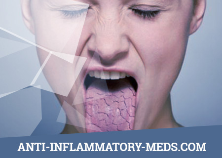
The basis for the diagnosis of Sjogren’s syndrome is a statement of two of the following three criteria in a patient: xerophthalmia, xerostomia, the presence of RA or other diffuse connective tissue disease. Very rarely, chronic aggressive hepatitis, primary biliary cirrhosis, or sarcoidosis can be a concomitant disease. Errors in the recognition of xerophthalmia and xerostomia are frequent, however. Sometimes patients exaggeratedly describe their sensations as dry eyes and mouth, and in such cases, focused questioning and examination immediately make sure that there is abundant secretion of saliva and tears. Very useful in this regard are simple questions about the possibility of swallowing dry food without washing down with liquid, about the presence of tears during heavy experiences or cutting onions, etc. The manifestations of true xerostomia and xerophthalmia are outlined in the Clinical Picture section.
The question of Sjogren’s syndrome arises not only with complaints of patients about dryness, but also with an increase in the salivary glands, especially the parotid glands. In such situations, the doctor must bear in mind that their increase is characteristic, in addition to the dry syndrome, also of tumor pathology (primary benign and malignant tumors of the salivary glands, lymphosarcoma, Waldenstrom macroglobulinemia), inflammatory diseases (sarcoidosis, acute bacterial and viral infections of the glands, chronic sialadenitis , tuberculosis, syphilis, actinomycosis, histoplasmosis, hookworm) and a number of other diseases (allergies to iodine, lead and copper compounds, salivary duct stones, diabetes mellitus, cirro PS liver, hyperlipidemia, starvation).
Tumors should especially be thought of with one-sided progressive enlargement of the gland, its high density and the absence of other signs of dry syndrome. In favor of Sjogren’s syndrome, fluctuations in the size of the glands, the absence of pain in them, a chronic enlargement of the glands without involving the surrounding tissues in the process indicate.
It must be remembered that an increase in salivary and lacrimal glands can occur with lymphosarcoma, lymphocytic leukemia and sarcoidosis, but dry syndrome among these diseases is possible only with sarcoidosis. In doubtful cases, a biopsy of one of the small salivary glands can be used to confirm the diagnosis of Sjogren’s syndrome. A large gland biopsy is warranted only if a tumor is suspected. Most of the other diseases listed above with a correct assessment of their clinical picture and medical history of patients can be quite clearly differentiated from Sjogren’s syndrome (primarily due to the absence of xerophthalmia and xerostomia).
For the correct diagnostic generalization in rare severe cases of primary Sjogren’s syndrome, it is necessary to take into account that the following syndromes, in addition to xerostomia and xerophthalmia, can be the clinical symptoms of this disease: vasculitis (panarteritis, necrotizing arteritis); hyperglobulinemic purpura; lymphoproliferation and related diseases; hepatosplenomegaly; an increase in the parotid salivary glands; fibrosing alveolitis and signs of obstructive airway disease; Raynaud’s syndrome; renal tubular acidosis; glomerulonephritis; polyneuritis; anemia, leukopenia, thrombocytopenia; myositis.
The correct idea of the possibility of these syndromes is important for the correct diagnosis within a single nosological unit, without resorting to the assumption of a combination of several diseases. It should be noted once again that the above systemic pathology, especially in the aggregate, is relatively rare in primary Sjogren’s syndrome. To an even lesser extent, it is characteristic of patients with secondary Sjogren’s syndrome (if we take into account clinical symptoms that do not serve as a manifestation of a concomitant connective tissue disease).
Of great importance for the diagnosis of Sjogren’s syndrome is the detection in almost all patients of the Russian Federation, and in a number of patients in the highest titers possible. In the primary dry syndrome, antibodies to Ro and La antigens are often found.
Treatment of Sjogren’s Syndrome
The treatment of patients with Sjogren’s syndrome usually consists in treating both the manifestations of the dry syndrome itself and the concomitant systemic disease (RA, SLE, etc.). The treatment of concomitant diseases is carried out without any fundamental features, however, doctors need to keep in mind a slightly greater tendency of patients with Sjogren’s syndrome to allergic reactions to drugs. Therefore, the purpose of the latter should be strictly justified. As a rule, after discharge from the hospital, patients should be under the supervision of a rheumatologist, ophthalmologist and dentist. It is necessary to pay special attention to a possible increase in the size of the salivary glands and the appearance of signs of lymphoproliferation in order to conduct an appropriate oncological examination.
For the treatment of dry syndrome as such, symptomatic agents are mainly used that significantly improve the condition and well-being of many patients. At the same time, it is undesirable to prescribe antihistamines and antidepressants, which can significantly increase the dryness of the mucous membranes.
With dry keratoconjunctivitis, at least half of the patients receive significant relief from instillation of “artificial tears” in the eye, the main component of which is usually a 0.5% methylcellulose solution. Since, in addition to dryness, in some cases, the accumulation of viscous mucus causes great inconvenience, such patients should use a 5-10% solution of the mucolytic drug acetylcysteine in the form of eye drops. The frequency of instillation of these drugs is determined individually. Some authors note the effectiveness of soft contact lenses that slow the evaporation of tears from the eyes. While maintaining a small lacrimation, the closure of the nasolacrimal canals by electrocoagulation is successfully used. In cases of secondary bacterial or fungal infection of the eyes, vigorous treatment with appropriate antibiotics is indicated.
Xerostomia, especially with great severity, is difficult to treat. The jelly-like lubricants (lubricants) used for this purpose were ineffective, and in some cases increased the feeling of dry mouth. Therefore, the use of water or other fluids remains the best method of controlling xerostomia, which makes chewing easier and the oral cavity free of food residues. Careful dental care and regular dental consultations are necessary for the active treatment of the first symptoms of complications (gingivitis, caries, thrush, etc.). With rare purulent mumps, which cannot be treated with antibiotics, as well as with complete blockage of the salivary duct with a stone, surgical intervention is necessary.
Dry nose decreases when irrigated with isotonic sodium chloride solution. Intranasal administration of lubricants containing various oils is undesirable, due to the risk of developing lipoid pneumonia. Dry skin rarely requires special treatment, usually emollient creams are quite enough. When the vagina is dry, softening gels are prescribed.
Attempts have also been made to reduce the dryness of the mucous membranes with the help of not local, but general effects, in particular bromhexine in a daily dose of 24-48 mg. A controlled randomized study showed that the administration of this drug led to an increase in the secretion of tears, but not saliva, which was the main calculation.
In patients with primary Sjogren’s syndrome, in addition to the methods of local treatment of dry mucous membranes discussed above, if necessary, drugs of a different action are also used: alkali for renal tubular acidosis, NSAIDs for arthralgia and arthritis, nifedipine for Raynaud’s syndrome. In cases of severe myositis, rare hemolytic anemia or severe systemic vasculitis with neurological complications, corticosteroids are indicated in moderate daily doses (20-40 mg of prednisolone) with the usual gradual dose reduction to the minimum maintenance and subsequent withdrawal. With the development of lymphoproliferative syndrome (enlarged lymph nodes and spleen, often in combination with an increase in the size of the salivary glands and cryoglobulinemic purpura and pulmonary infiltration), corticosteroids are also prescribed (starting with 30-40 mg of prednisolone per day), often in combination with cyclophosphamide or azathioprine. Local radiotherapy of enlarged salivary glands with Sjogren’s syndrome is contraindicated in connection with the risk of lymphosarcoma. Prescribing prednisone only for an increase in the salivary or lacrimal glands is also not recommended, since the dryness of the mucous membranes as a result of this treatment does not decrease, despite a possible reduction in the size of the spleen. There are reports of positive treatment results for patients with Sjogren’s syndrome only with cyclophosphamide, but they are few in number and therefore need confirmation.
In recent years, evidence has emerged of the successful use in severe cases of Sjogren’s syndrome of pulse therapy with methylfednisolone (for 3 consecutive days at 1000 mg / day slowly intravenously) or methylprednisolone in combination with cyclophosphamide (the latter at a dose of 1000 mg is additionally administered on the first day of tulotherapy). As a result, the signs of xerostomia of 4 xerophthalmia decreased, clinical improvement of systemic manifestations and positive dynamics of immunological parameters were noted. The authors believe that pulse therapy can be considered the method of choice for such manifestations of Sjogren’s syndrome as vasculitis with ulcerative necrotic changes in the skin, polyneuritis, hyperglobulinemic (cryoglobulinemic) purpura, exudative polyserositis, cerebrovascularitis and autoimmune hemolytic anemia. According to the observations of the same authors, the results of pulse therapy increase even more when it is combined with plasmapheresis or hemosorption.