What is Reiter’s Syndrome (Disease)?
Reiter’s syndrome (illness) is a combined lesion of the genitourinary organs (usually in the form of nonspecific urethroprostatitis), joints (reactive oligo or monoarthritis) and the eye (conjunctivitis), developing simultaneously or sequentially.
Reiter’s disease (Reiter’s syndrome) occurs when people have a chlamydial infection with a genetic predisposition. A major role in the development of Reiter’s disease is played by disorders in the immune system that have arisen in connection with the formation of a focus of chronic chlamydial inflammation. Men get sick 20 times more often than women.
Causes of Reiter’s Syndrome (Disease)
The most common causative agent of the disease is the gram-negative bacterium Chlamydia trachomatis. Chlamydiae are obligate intracellular parasites with sizes of 250-300 nm. Under adverse conditions (exposure to antibiotics, chemotherapeutic drugs, etc.), chlamydia can transform into L-forms, which have the least ability to antigenically stimulate immunocompetent cells and are capable of prolonged intracellular parasitization. All this contributes to the chronic course of infection. This is the most common sexually transmitted infection and is the cause of non-gonococcal urethritis in men in 60% of cases. Chlamydial urethritis has a relapsing or chronic course. In women, chlamydia cause chronic cervicitis, salpingitis, adnexitis, cystitis. Women suffering from these diseases are carriers of chlamydia, but they themselves rarely suffer from urogenic arthritis.
Chlamydia trachomatis is sexually and non-sexually transmitted (domestic infection) and is found intracellularly in the epithelium of the urethra, conjunctiva and cytoplasm of synovial cells.
Reiter’s syndrome can be caused by Shigella, Salmonella, Yersinia and develop after suffering enterocolitis. Some experts point out that ureoplasma can cause Reiter’s disease. The hereditary predisposition matters, the marker of which is the HLA histocompatibility antigen – B27 (In 75-95% of patients).
Pathogenesis during Reiter’s Syndrome (Disease)
During sexual infection in the genitourinary organs (urethra, prostate, cervical canal of the uterus), a focus of inflammation develops, from where chlamydia spread to various tissues, including articular. Then an autoallergy develops, on the severity of which the nature of the course of the disease depends.
There are 2 stages of the disease: the first is infectious, characterized by the presence of chlamydia in the urethra; the second is immunopathological, accompanied by the development of immunocomplex pathology with lesions of the synovia of the joints and conjunctiva.
Other microorganisms can cause urethro-oculo-synovial syndrome – yersinia, salmonella, shigella. It is proposed to distinguish two forms of the disease: sporadic (Reiter’s disease) (venereal infection precedes clinical manifestations, and the etiological factor is detected in 65-70% of patients) and epidemic or postenterocolitic (arthritis, urethritis, conjunctivitis is preceded by enterocolitis of various nature – dysenteric, yersiniosis, salmonellosis undifferentiated). Some authors call the post-enterocolitic form Reiter’s syndrome.
It is assumed that chlamydia, shigella, salmonella, yersinia, ureaplasma have certain antigenic structures that cause a peculiar immune response in the form of an urethro-oculo-synovial syndrome in genetically predisposed individuals.
With a disease duration of up to 6 months, it is defined as an acute course, up to 1 year – protracted, more than 1 year – chronic.
Symptoms of Reiter’s Syndrome (Disease)
Mostly young men aged 20-40 years (80% of cases) fall ill, less often – women, extremely rarely – children. The onset of the disease is most often manifested by lesions of the genitourinary organs (urethritis, cystitis, prostatitis) a few days (sometimes a month) after sexual infection or transferred enterocolitis. Urethritis is manifested by unpleasant sensations during urination, burning, itching, hyperemia around the external opening of the urethra, and scanty discharge from the urethra and vagina. Discharge is usually mucous. It should be noted that urethritis is not extremely pronounced, as, for example, with gonorrhea and can be manifested only by small mucopurulent discharge from the urethra or only dysuria in the morning. Perhaps even the absence of dysuric phenomena. In 30% of men, urogenital chlamydia is asymptomatic. Such patients often have only initial leukocyturia or an increase in the number of leukocytes in a Gram-stained smear taken from a tampon inserted to a depth of 1-2 cm into the front of the urethra.
Eye damage occurs soon after urethritis, more often manifested by conjunctivitis, less often iritis, iridocyclitis, uveitis, retinitis, retrobulbar neuritis, keratitis. It should be noted that conjunctivitis can be mild, last 1-2 days and go unnoticed.
The leading sign of the disease is joint damage, which develops after 1-1.5 months. after acute urogenital infection or its exacerbation. The most characteristic asymmetric arthritis involving the joints of the lower extremities – knee, ankle, metatarsophalangeal, interphalangeal. Joint pain intensifies at night and in the morning, the skin above them is hyperemic, and effusion appears. Characteristic is the “ladder” (from bottom to top) sequential involvement of the joints in a few days. It is for urogenic arthritis (Reiter’s disease) that periarticular edema of the whole finger and sausage-like finger configuration with bluish-crimson skin color, as well as pseudogout symptoms involving the joints of the big toe, are pathognomonic.
Quite often there is inflammation of the Achilles tendon, bursitis in the heel area, which is manifested by severe heel pain. Perhaps the rapid development of heel spurs.
Some patients may develop pain in the spine and develop sarcoileitis.
More than 50% of patients have a complete disappearance of articular symptoms, 30% have relapses of arthritis, in 20% arthritis acquires a chronic course with limited joint function and atrophy of adjacent muscles. Some patients develop flat feet as a result of damage to the tarsal joints. Joints of the upper extremities are rarely affected.
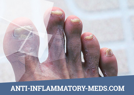
In 30-50% of patients, mucous membranes and skin are affected. Painful ulcers appear on the mucous membrane of the oral cavity and in the area of the glans penis, balanitis or balanoposthitis develops. Stomatitis, glossitis are characteristic. Skin lesions are characterized by the appearance of small red papules, sometimes erythematous spots. Extremely characteristic is keratoderma – draining glasses of hyperkeratosis on the background of hyperemia of the skin with cracks and peeling mainly in the feet and palms. Foci of hyperkeratosis can be observed on the skin of the forehead, trunk.
Possible painless enlargement of the lymph nodes, especially inguinal; 10-30% of patients have signs of heart damage (myocardial dystrophy, myocarditis), lung damage (focal pneumonia, pleurisy), nervous system (polyneuritis), kidneys (nephritis, renal amyloidosis), prolonged low-grade body temperature..
Diagnosis of Reiter’s Syndrome (Disease)
Diagnostic Criteria for Reiter’s Disease
- The presence of a chronological relationship between a genitourinary or intestinal infection and the development of symptoms of arthritis and / or conjunctivitis, as well as lesions of the skin and mucous membranes.
- The young age of the sick.
- Acute asymmetric arthritis mainly of the joints of the lower extremities (especially the joints of the toes) with enthesopathies and calcaneal bursitis.
- Symptoms of the inflammatory process in the genitourinary tract and the detection of chlamydia (in 80-90% of cases) in scrapings of the epithelium of the urethra or cervical canal.
In the absence of chlamydia in the scrapings of the urethra of the cervical uterus, seronegative arthritis can be regarded as chlamydial if there are signs of inflammation of the urogenital sphere, and chlamydial antibodies are found in the blood serum in a titer of 1:32 or more.
Laboratory data for Reiter’s disease
- General blood test: signs of slight hypochromic anemia, moderate leukocytosis, increased ESR.
- Urinalysis. Leukocyturia in samples according to Nechiporenko, Addis-Kakovsky, in a three-glass sample, leukocytes are mainly in the first portion of urine.
- The study of prostate secretion – more than 10 leukocytes in the field of view, a decrease in the number of lecithin grains.
- Biochemical analysis of blood: increased levels of alpha2- and beta-globulins, sialic acids, fibrin, seromucoid, the appearance of PSA. RF is negative.
- Detection of chlamydial infection. A cytological examination of scrapings of the mucous membrane of the urethra, cervical canal, conjunctiva, as well as sperm and prostate juice is performed. Scraping is done with a Volkman spoon or special sterile brushes. Chlamydia tropism to the cylindrical epithelium of the genitourinary tract, so scraping should be done so that these cells are present. The contents of the scraping is distributed on a glass slide, fixed with methanol and stained according to Romanovsky-Giemsa, chlamydia are detected in the form of intracellular inclusions. To prepare preparations from synovial fluid, it is centrifuged for 10 minutes and the precipitate is washed with Hanks solution. The suspension is placed on glass and stained according to Romanovsky-Giemsa. This color has a low reliability (5-15%). Chlamydia luminescent bacterioscopy is more promising when staining the drug with monoclonal antibodies labeled with fluorochrome, i.e. method of fluorescent antibodies. The sensitivity of the method is 95%. Chlamydial antibodies in the blood can be detected using the complement binding reaction, indirect hemagglutination reaction and immunofluorescence analysis. Serological tests are auxiliary, since in 50% of patients, due to low immunogenicity, antibodies are not detected. The best method for diagnosing chlamydial infection is DNA diagnostics using the polymerase chain reaction. The test has species specificity comparable to the cell culture method and has a sensitivity of 10 DNA molecules of any type of chlamydia. If you suspect an enterocolitic form of Reiter’s disease using bacteriological and serological methods, salmonella, shigellosis, and yersiniosis infections should be excluded.
- Synovial fluid research – inflammatory type changes: a mucin clot is loose, the number of leukocytes is 10-50 × 109 / l, neutrophils make up more than 70%, cytophagocytic macrophages, chlamydial antigens and antibodies are detected, a high level of complement, rheumatoid factor is not determined.
- Carriage of HLA B27 is revealed
Instrumental research
X-ray examination of the joints reveals asymmetric periarticular osteoporosis, asymmetric narrowing of the joint spaces, with a long course – erosive-destructive changes, due to periostitis – calcaneal spurs and isolated spurs on the body of one or two vertebrae; pathognomonic spurs of the metacarpal bones and their erosion periostitis of the calcaneus and phalanges of the toes, asymmetric erosion of the metatarsophalangeal joints, in 30-50% of patients – signs of sacroileitis, often unilateral.
Prevention of Reiter Syndrome (Disease)
Have one reliable sexual partner or use a condom during casual sexual intercourse. Begin sex education from childhood.

